Overview
Microtubules are cytoskeletal filaments that determine spatial organization, provide tracks for intracellular trafficking, generate the mitotic spindle for cell division, and comprise the axoneme of primary and motile cilia. Our recent work falls into three broad areas of microtubule biology:
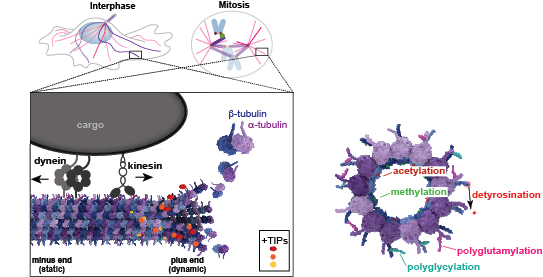
“The Tubulin Code“
All microtubules are polymerized from the same building block, the protein tubulin, yet cells can generate functionally diverse microtubules that play specific roles in spatial organization, directional transport, and force generation. Microtubule diversity can arise from sequence differences across the tubulin isotypes encoded by the human genome and from post-translational modifications to tubulin subunits within a microtubule. This diversity has been proposed to generate a “Tubulin Code” that is recognized by cellular factors, i.e., “readers” that carry out specific microtubule-based functions.
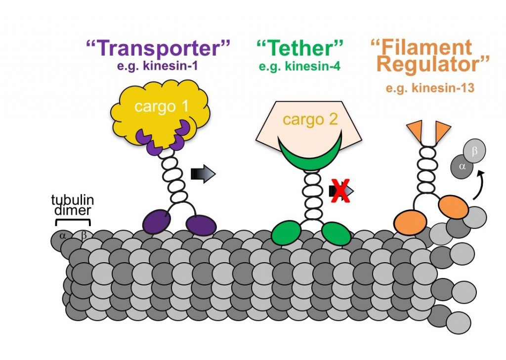
“Kinesin mechanics and function“
All kinesin proteins contain a kinesin motor domain that binds both ATP and microtubules. Sequence differences between kinesin motors allow them to utilize ATP and interact with microtubules for unique biological outputs. Some kinesins are highly processive and capable of long-distance transport whereas some are immotile and may serve as tethers for signaling complexes and others
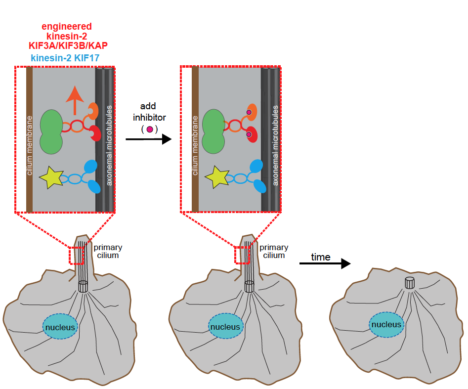
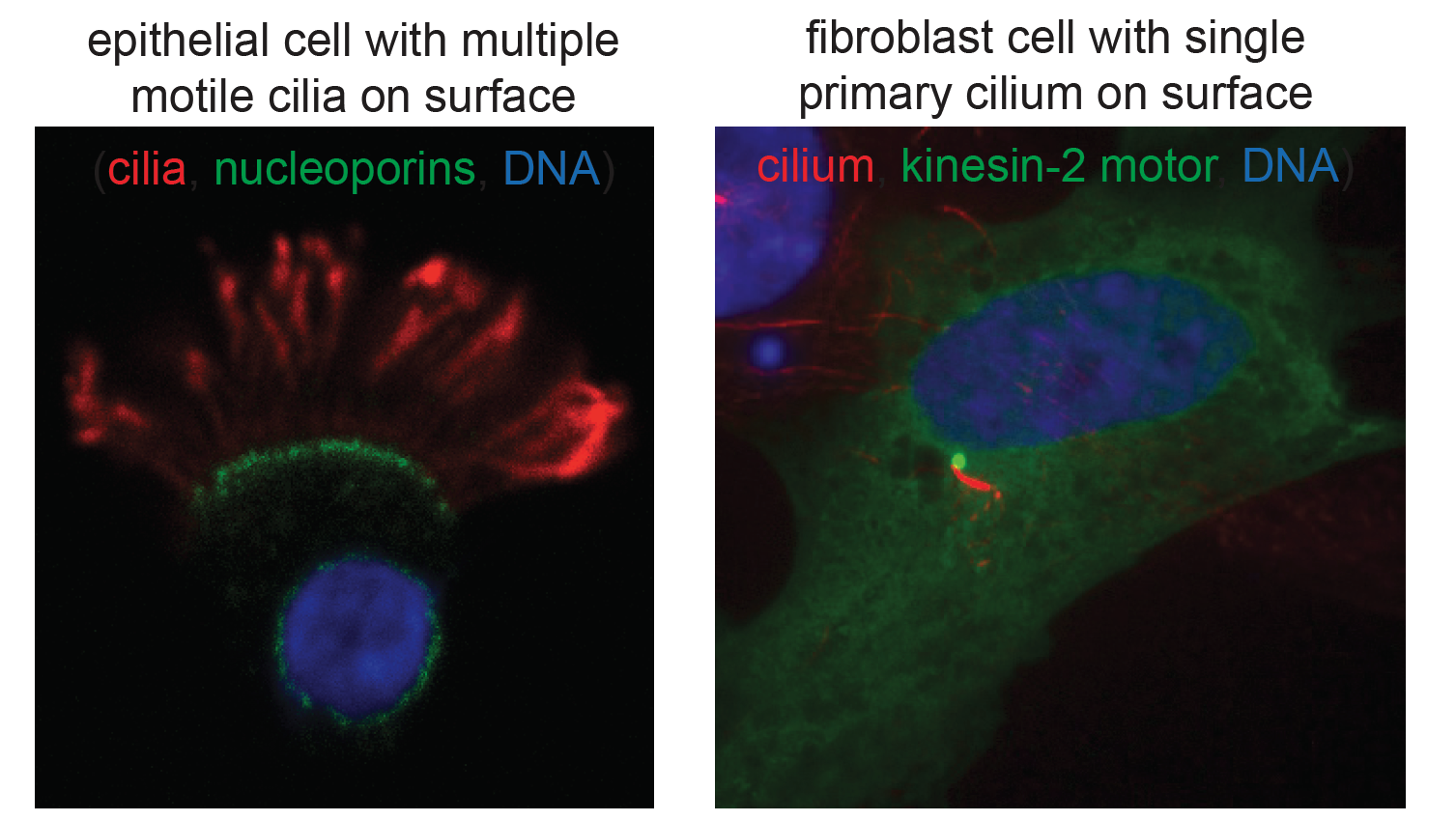
“Primary cilia as sensory organelles“
Cilia (and eukaryotic flagella) are microtubule-based organelles that protrude from the surface of nearly all cells in the human body and play important roles during development and normal adult physiology. The assembly, maintenance and function of cilia depend on microtubule-based transport by members of the kinesin-2 family.